. 2015 Feb;110(1):23–47. doi: 10.1590/0074-02760140266

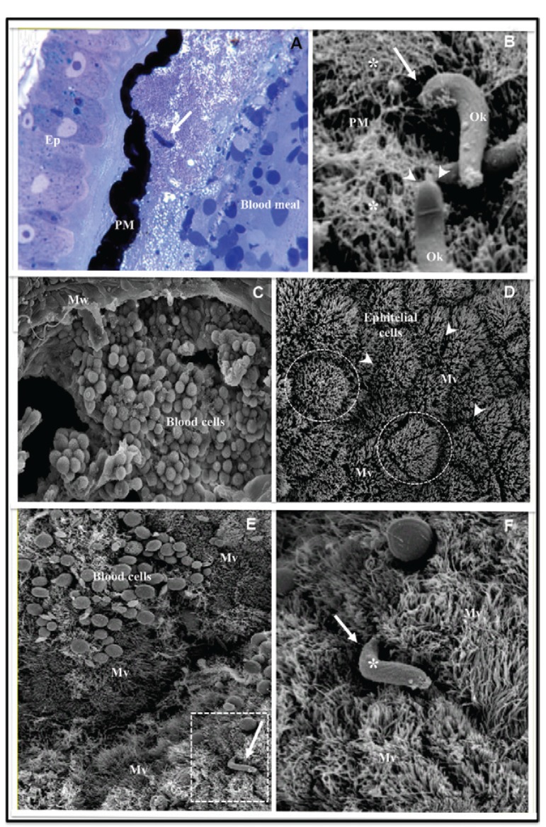
Fig. 1: histology (A) and scanning electron microscopy (SEM) (B-F) of Anopheles aquasalis midguts after a Plasmodium vivax infective blood meal. A: historesin section of a midgut stained with Giemsa. The peritrophic matrix (PM) sturdily stained in black is separating the midgut epithelium (Ep) from the blood meal. Note an ookinete (Ok) (arrow) close to the PM; B: SEM of an opened midgut showing two Oks over the PM. Observe the fibrous aspect (asterisks) of the internal side of the PM. One Ok is crossing the PM throughout the fibre layer (large arrow). Another Ok is showing details of its anterior extremity (arrowheads); C: small magnification of an opened midgut showing the blood meal containing the numerous blood cells. Note a portion of the midgut wall (Mw); D: large magnification of an opened midgut showing details of the epithelial cells. The epithelial cells have polygonal shapes (circles) and their surfaces are covered by microvilli (Mv). Note the clefts (arrowheads) among the epithelial cells; E: small magnification of an opened midgut with blood cells of the blood meal. Note inside the square area one Ok (arrow) penetrating the Ep Mv; F: large magnification of the square area of E in the Figure showing details of the Ok penetration. Note the Ok (asterisk) extremity inserted in a cleft (asterisk) among the epithelial cell Mv.

Fig. 1: