
Figure 1. MRI lesions characteristic of neuromyelitis optica spectrum disorder.
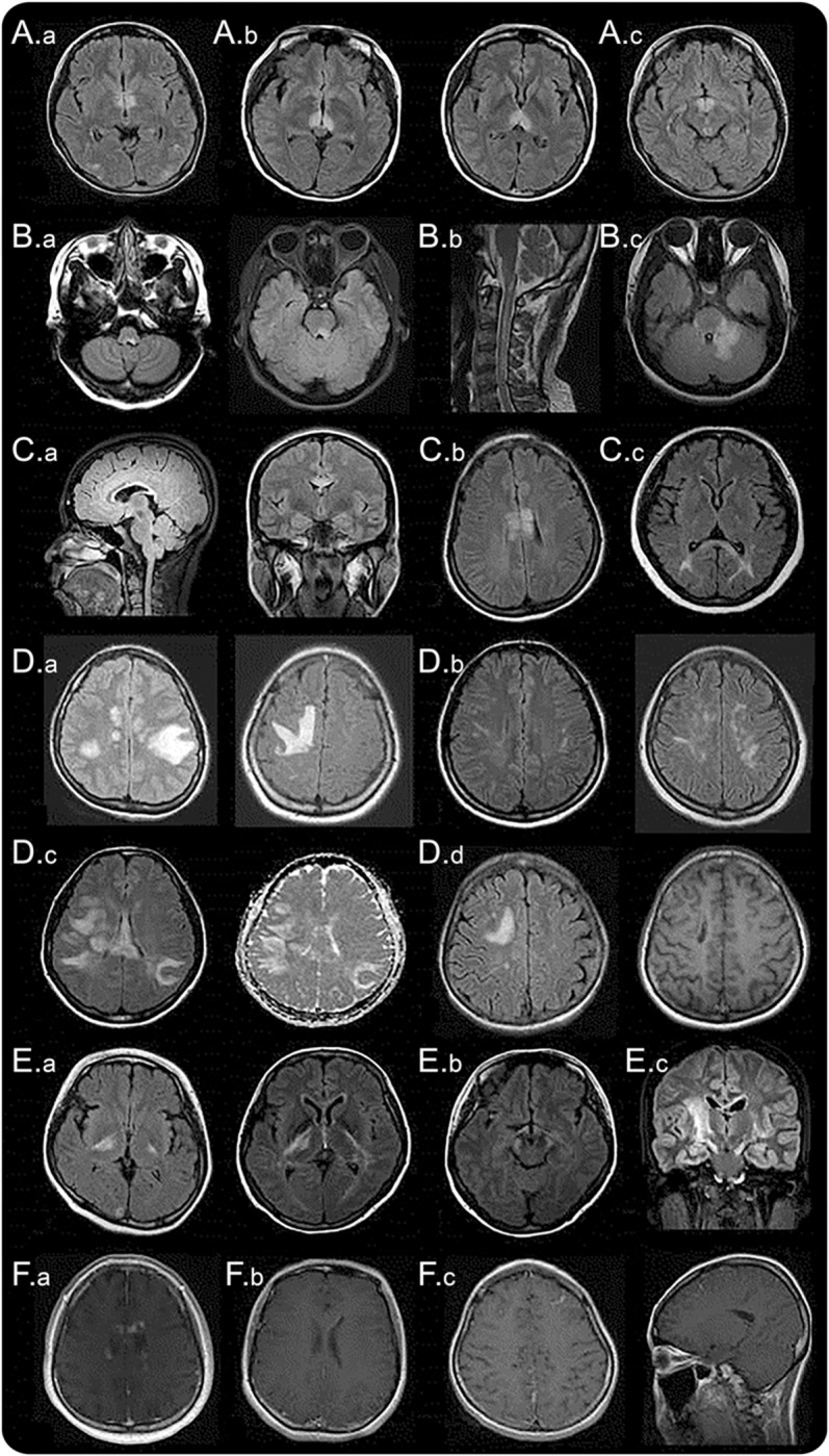
Diencephalic lesions surrounding (A.a) the third ventricles and cerebral aqueduct, (A.b) which include thalamus, hypothalamus, and (A.c) anterior border of the midbrain. (B.a) Dorsal brainstem lesion adjacent to the fourth ventricle, (B.b) linear medullary lesion that is contiguous with cervical cord lesion, (B.c) edematous and extensive dorsal brainstem lesion involving the cerebellar peduncle. (C.a) Callosal lesion immediately next to the lateral ventricle, following the ependymal lining, (C.b) “marbled pattern” callosal lesion, (C.c) “arch bridge pattern” callosal lesion. (D.a) Tumefactive hemispheric white matter lesions, (D.b) a long spindle-like or radial-shape lesion following white matter tracts, (D.c) extensive and confluent hemispheric lesions show increased diffusivity on apparent diffusion coefficient maps suggesting vasogenic edema, (D.d) hemispheric lesions in the chronic phase showing cystic-like cavitary changes. (E.a) Corticospinal tract lesions involving the posterior limb of the internal capsule and (E.b) cerebral peduncle of the midbrain, (E.c) longitudinally extensive lesion following the pyramidal tract. (F.a) Cloud-like enhancement, (F.b) linear enhancement of the ependymal surface of the lateral ventricles, (F.c) meningeal enhancement.