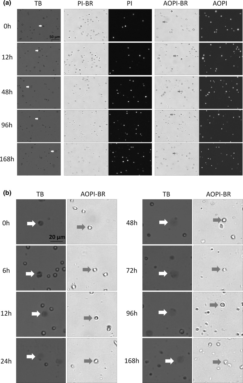
Fig. 1.
Bright-field and FL images of PI, AO/PI, and TB stained naturally-dying Jurkat cells. a Samples were measured at time points 0, 6, 12, 24, 48, 72, 96, and 168 h post incubation at room temperature. b Higher magnification of a showing TB time-course study. Large, dim, and diffused shaped cells in the TB and bright-field images are shown with red and green arrows, respectively. (Color figure online)

