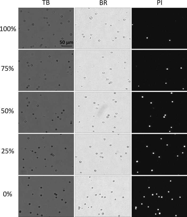
Fig. 6.
Bright-field and fluorescent images of TB and PI-stained heat-killed Jurkat cells at various viability percentages. The heat-killed and fresh Jurkat cells were mixed to produce five different viability percentages (0–100 %). In these heat-killed Jurkat samples the TB images showed a clear distinction between live and dead cells; where the dead cells exhibited a tight dark and well-defined morphology. Similarly, the dead cells in the bright-field images maintained an easily detectable and sharp cellular membrane

