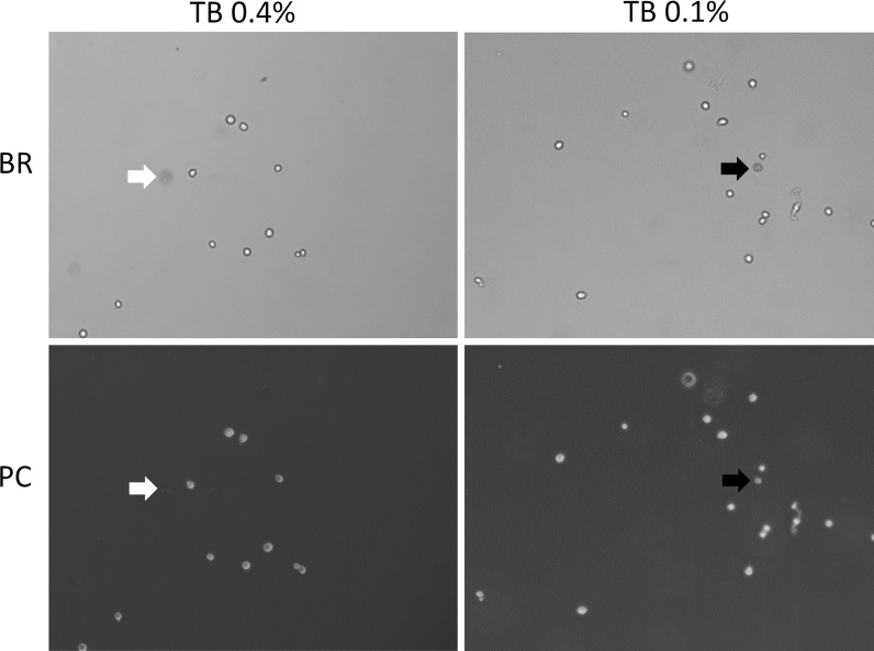
Fig. 8.
Phase contrast and BR microscopy images. Both phase contrast and bright-field images were captured using digital camera on a light microscope. The red arrow indicated the dim diffused cell in 0.4 % TB-stained Jurkat cells, where it was not observed in the phase contrast image. The green arrow indicated the dark dead cell in 0.1 % TB-stained Jurkat cells, where it was also observed in the phase contrast image. (Color figure online)

