Figure 2.

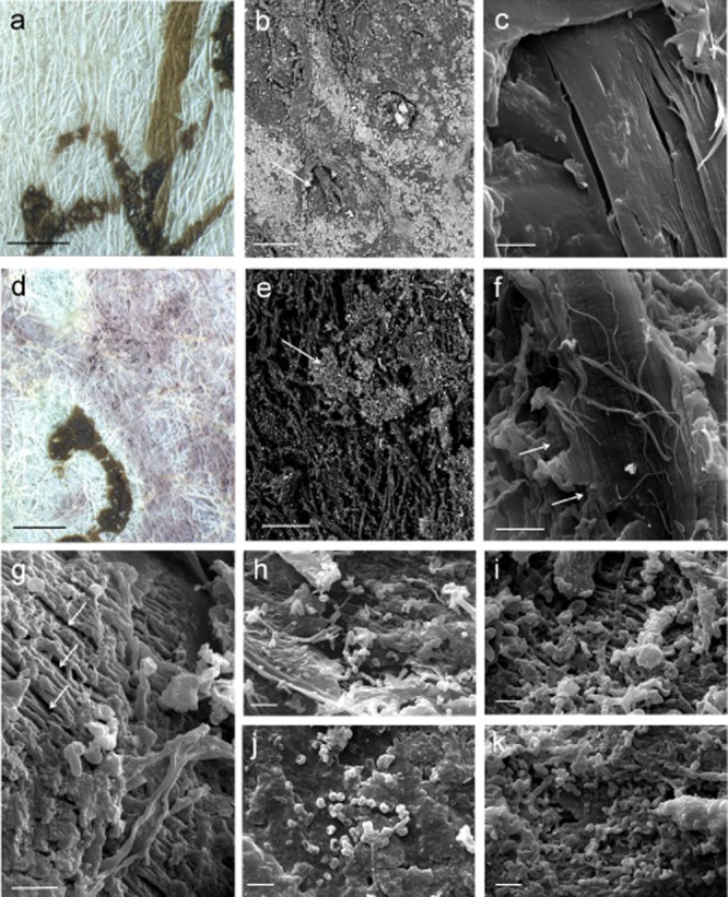
Comparison of healthy and damaged parchment:A. Detail of the surface of unstained Ve-21 parchment obtained with a Leica MZ16 stereomicroscope, the brown sign is the ink of the writing (bar = 1 mm).B. SEM image of a healthy Ve-21 parchment sample. The picture was obtained at variable pressure (50Pa) with a backscattered electron detector on not metallized material. The arrow indicates a hair follicle and a portion of hair. The surface is covered with mineral particles appearing lighter according to their higher atomic number (bar = 100 μm).C. High-vacuum secondary electrons SEM image on gold sputtered samples of a healthy Ve-21 parchment sample. The collagen fibres appear smooth and homogeneous (bar = 2 μm).D. Detail of the surface of purple stained Ve-21 parchment obtained with a Leica MZ16 stereomicroscope, the brown sign is the ink of the writing (bar = 1 mm).E. SEM image of biodeteriorated Ve-21 parchment sample. The picture was obtained at variable pressure (50Pa) with a backscattered electrons detector on not metallized material. The arrow indicates the original surface which was covered with mineral particles appearing lighter according to their higher atomic number (bar = 100 μm).F. High-vacuum secondary electrons SEM image on gold sputtered samples of a biodeteriorated Ve-21 parchment sample. Fibrils of collagen can still be appreciated (arrow), but a general breakdown of the material is present (bar = 2 μm).G. High-vacuum secondary electrons SEM image on gold sputtered samples of a biodeteriorated Ve-3 parchment sample. It can be seen the formation of cracks between the bundles of fibrils, and disintegration of the matrix that holds together the fibrils (arrows) (bar = 2 μm).H–I. Chains of bacterial spores and filaments on sample 148D consistent with Actinomycetales order (a, bar = 3 μm; b, bar = 1 μm).J. Bacterial spores (about 600 nm diameter) on sample Ve-21 (bar = 1 μm).K. Chains of bacterial spores and filaments on sample Ve-21 consistent with Actinomycetales order (bar = 1 μm).
