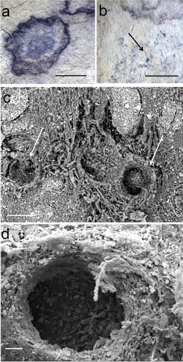
Figure 3.

Purple stains from sample To800:A–B. Details of the stains obtained with a Leica MZ16 stereomicroscope (bar = 2 mm); The arrow in panel B indicates spots corresponding to Chaetomium sp. peritecia, immersed in the substrate (bar = 5 mm).C. SEM picture obtained at variable pressure (50Pa) with a backscattered electrons detector on not metallized material. Chaetomium fruiting structures immersed in parchment (bar = 100 μm).D. HV SE SEM image on a gold sputtered sample of the fractured fungal peritecia immersed in parchment (bar = 20 μm). The hole is produced by the fungus and consists of the inner walls of the fruiting body. On the on the bottom of the crater there are some ascospores.
