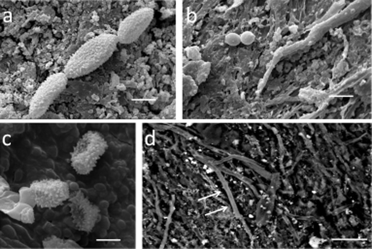
Figure 4.

SEM imaging of fungi on parchment samples:A. HV-SEM on gold sputtered samples, chains of verrucolose conidia and consistent with some species of Cladosporium and Toxicocladosporium were observed on MS-492 sample (bar = 5 μm).B. HV-SEM on gold sputtered samples, obovoid conidia with a truncate base, consistent with Phialosimplex, were documented on Ve-3 (bar = 10 μm).C. HV-SEM on gold sputtered samples, echinated conidia and conidiophora resembling a species of Penicillium were documented on samples Ve-3 and Ve-21 (bar = 2 μm).D. VP-BSD-SEM, surface of the parchment sample Bo-Arch with fungal hyphae (arrows) growing adherent to collagen fibres (bar = 20 μm).
