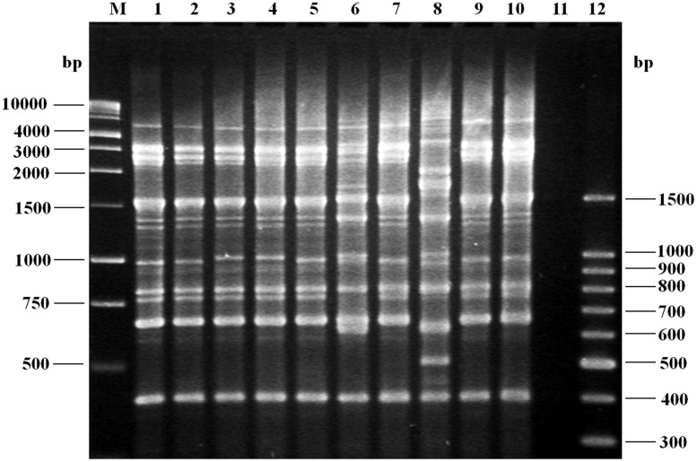
FIGURE 3.
Agarose gel (1.5%) electrophoresis showing the results of polymerase chain reaction (PCR) amplification of representative fingerprint patterns for the REP-PCR of Vibrio parahaemolyticus strains of serotype O3:K6 (tdh+, toxRS/new+, orf8+, and trh-), isolated from 2004 to 2013. Lane M, 1 kb molecular size markers; lane 1, clinical isolate (2004, O3:K6); lane 2, clinical isolate (2006, O3:K6); lane 3, clinical isolate (2008, O3:K6); lane 4, clinical isolate (2010, O3:K6); lane 5, clinical isolate (2011, O3:K6); lane 6, shrimp isolate (2011, O3:KUT, tdh+, toxRS/new+, orf8+, and trh-); lane 7, clinical isolate (2012, O3:K6); lane 8, clinical isolate (2012, O1:K20, tdh-, toxRS/new-, orf8-, and trh-); lane 9, clinical isolate (2013, O3:K6); lane 10, control strain V. parahaemolyticus RIMD 2210633 (tdh+, toxRS/new+, orf8+, and trh-); lane 11, negative control (no DNA template added to the rep-PCR reaction); lane 12, molecular size markers.

