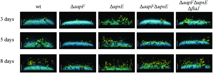
FIGURE 4.
The loss of distinct type IV pili results in different biofilm structures. Side view of S. acidocaldarius biofilms after 3, 5, and 8 days of incubation. The blue channel represents DAPI staining. The green channel shows fluorescent labeled lectin ConA, which binds to glucose and mannose residues. The lectin IB4 binds galactosyl residues and is shown in yellow. Scale bar = 40 μm (Henche et al., 2012b). Image courtesy of Sonja Albers, University of Freiburg.

