
Figure 5.
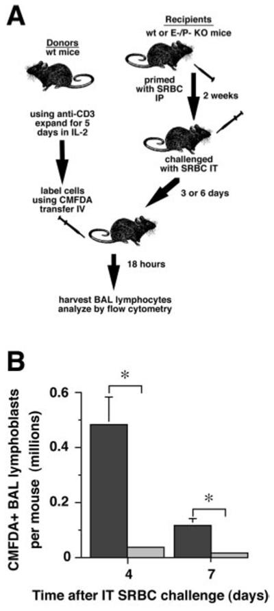
Recruitment of T lymphoblasts into lungs of E-P- mice is reduced. A. Experimental protocol. Lymph node T cells from normal wt mice were activated using immobilized anti-CD3 mAb and their numbers expanded in IL-2 to produce T lymphoblasts, as previously described (16). After 5 days of expansion, T lymphoblasts were labeled with CMFDA and transferred by tail vein injection into primed recipients consisting of either wt or E-P- mice which had been IT Ag challenged three or six days earlier. BAL lymphocytes were analyzed 18 hours later by flow cytometry. B. Results. Dark bars, wt recipients; light bars, E-P- recipients. Data are mean ± SD of individual recipient mice (wt, 3 mice, E-P-, 2 mice); *, p<0.05, unpaired t test.