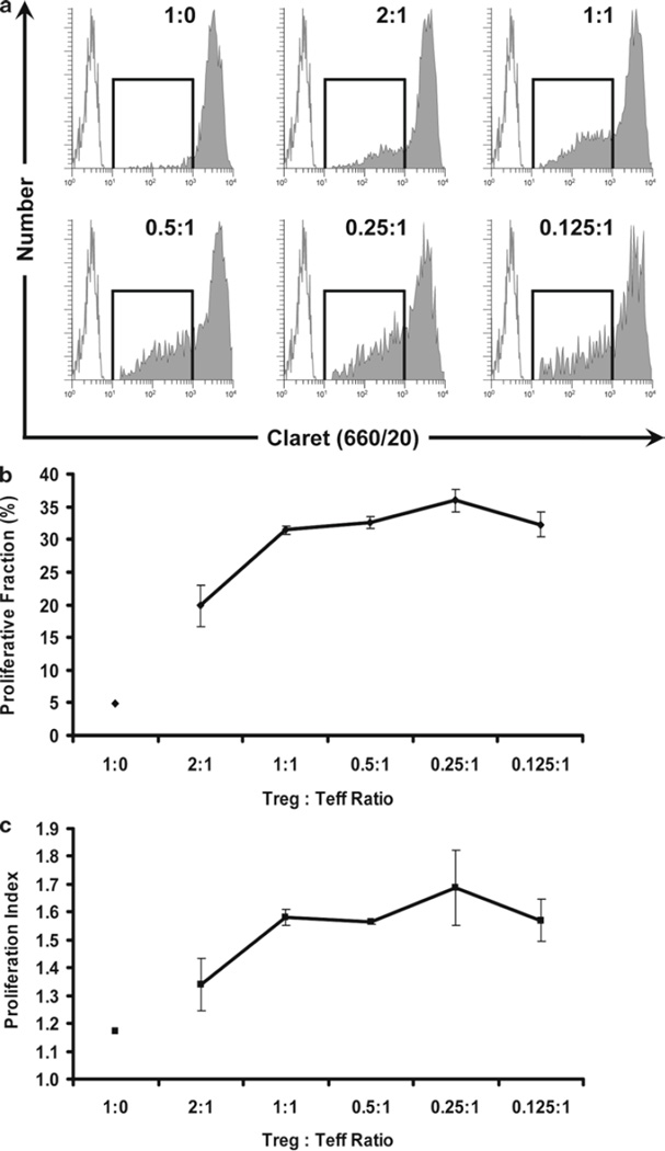
Fig. 10.
Enhanced Proliferation of Treg cells at low Treg:Teff ratios. The use of a two-dye system allows for the discrimination of Treg and Teff from accessory cells and from each other, and the simultaneous measurement of proliferation in both subsets. In the same samples as shown in Fig. 9, the proliferation of CellVue Claret-stained Treg cells was monitored at different Treg:Teff ratios. Treg are generally anergic and, as expected, did not proliferate when incubated with anti-CD3, anti-CD28, and accessory cells in the absence of Teff cells (Treg:Teff ratio of 1:0). However, as the proportion of Teff in the cultures was increased (i.e., as the Treg:Teff ratio decreased), the extent of Treg proliferation also increased. Proliferative fraction (%P) and Proliferative Index (PI) were determined as described in Fig. 8 and Subheading 3.4.5. Data points in (b) and (c) represent the mean ± 1 standard deviation of triplicate samples. (a) Representative proliferation profiles for Treg at varying Treg:Teff ratios showing increasing proliferation at lower ratios (increased proportion of Teff in cultures) (filled histograms). Unstained, unstimulated cells (unfilled histograms) are overlaid for reference. (b) Effect of Treg:Teff ratio on Proliferative Fraction of Treg. (c) Effect of Treg:Teff ratio on Proliferative Index of Treg.

