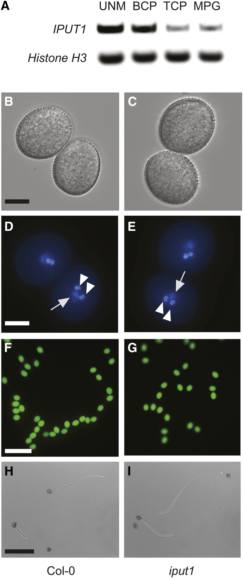
Figure 5.
iput1 Pollen Develops Normally.
(A) RT-PCR of IPUT1 and Histone H3 (control) in the uninucleate microspore (UNM), bicellular pollen (BCP), tricellular pollen (TCP), and mature pollen grains (MPG). RT-PCRs used pooled spores from two biological replicates.
(B), (D), (F), and (H) Pollen from wild-type plants.
(C), (E), (G), and (I) Pollen from iput1-1/+ plants. Phenotype of wild type and iput1 pollen is indistinguishable.
Light microscopy of mature pollen grains ([A] and [B]); fluorescence microscopy after DAPI staining ([D] and [E]) and fluorescein diacetate staining ([F] and [G]). Arrows indicate the diffusely stained vegetative nucleus; arrowheads indicate the densely stained sperm cell nuclei. Light microscopy of germinated pollen tubes, 6 h after germination ([H] and [I]). Bars = 10 μm in (B) to (F) and 100 μm in (G) to (I).

