Figure 2.
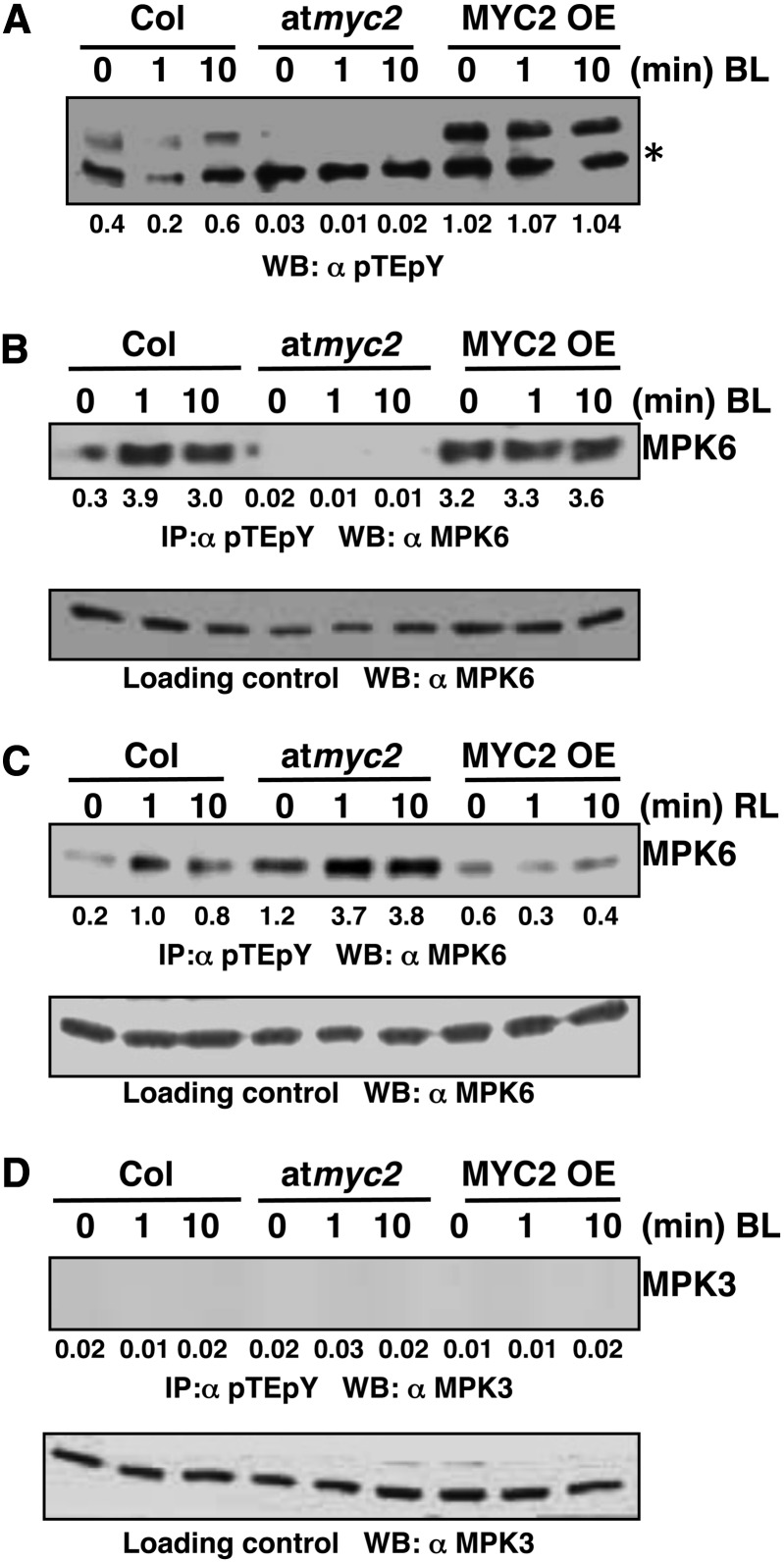
MPK6 Activation in BL Is MYC2 Dependent.
(A) Immunoblot analysis using pTEpY antibody of 50 μg of total protein from dark-grown Col, myc2, and MYC2 OE lines exposed to BL (30 μmol m−2 s−1) for 0, 1, or 10 min. Upper bands indicate total active MAPKs, and lower bands (asterisk) indicate cross-reacting band. Values below the panel indicate quantification of each band with respect to the loading control (lower cross-reacting band).
(B) Immunoprecipitation assays using 200 μg of total protein from dark-grown Col, myc2, and MYC2OE lines exposed to BL (30 μmol m−2 s−1) for 0, 1, or 10 min. The MPK6 activation was analyzed by immunoprecipitation with pTEpY antibody followed by immunoblot analysis with anti-MPK6. Lower panel shows immunoblot with MPK6 antibody using 20 μg of total protein as the loading control. Values below the panel indicate quantification of each band with respect to MPK6, the loading control.
(C) Immunoprecipitation assay of dark-grown Col, myc2, and MYC2OE lines exposed to RL (60 μmol m−2 s−1) for 0, 1, or 10 min. The specific MPK6 activation was analyzed by immunoprecipitation with pTEpY antibody followed by immunoblot analysis with anti-MPK6. The lower panel shows an immunoblot with MPK6 antibody using 20 μg of total protein as loading control. Values below the panel indicate quantification of each band with respect to MPK6, the loading control.
(D) MPK3 is not activated in BL. Immunoblot probed with anti-MPK3 after immunoprecipitation with pTEpY antibody acts as negative control. Lower panel shows immunoblot analysis with anti-MPK3 using 20 μg of total protein as loading control.

