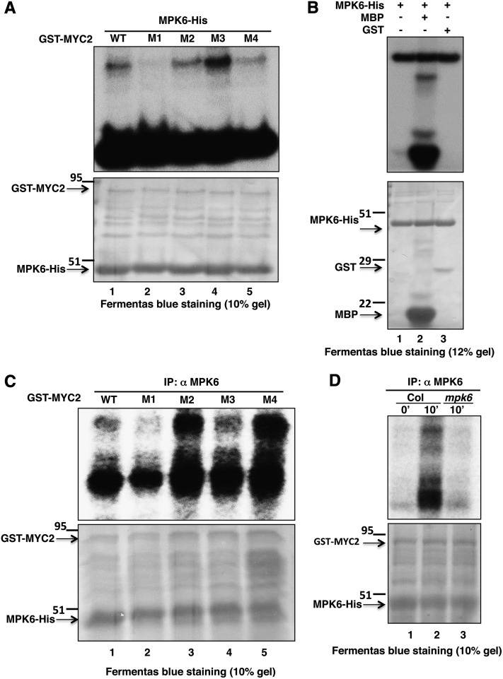
Figure 4.
MPK6 Phosphorylates MYC2 in BL.
(A) In vitro kinase assay using bacterially expressed MPK6-His and GST-MYC2 (wild-type protein and different site-directed mutagenized versions, i.e., M1-M4 of MYC2 protein, lanes 1 to 5, respectively). GST-MYC2 M1 protein has Ser-123 replaced with Ala, βM2 has Thr-287 replaced with Ala, M3 has Thr-328 replaced with Ala, and M4 has Ser-330 replaced with Ala. Lower panel shows PAGE-blue stained gel showing position of different proteins.
(B) Affinity-purified proteins (MPK6-His and GST) were expressed in Escherichia coli. MPK6-His was incubated alone or in combination with GST or MBP in the kinase reaction mixture to check their phosphorylation by MPK6. The plus and minus signs indicate the presence and absence of proteins, respectively. Aliquots of the samples were separated by SDS-PAGE and subjected to autoradiography. Lower panel shows the PAGE-blue stained gel with the position of different proteins indicated (arrows).
(C) In vitro phosphorylation assay using plant protein. Arabidopsis wild-type (Col) seedlings grown in dark and then exposed to BL (30 μmol m−2 s−1) for 10 min. Immunoprecipitation of crude protein (300 μg) with anti-MPK6 followed by incubation with GST-MYC2 as substrate (wild type or mutated, lanes 1 to 5, respectively) in the kinase reaction mixture to determine phosphorylation activity. PAGE-blue stained gel, with the position of different proteins indicated (arrows).
(D) Immunoprecipitation of MPK6 from Arabidopsis wild-type (Col) seedlings grown in constant darkness (lane 1) and Col and mpk6 seedlings grown in darkness for 6 d and then exposed to BL (30 μmol m−2 s−1) for 10 min (lanes 2 and 3, respectively), followed by incubation with GST-MYC2 as substrate in the kinase reaction mixture to determine phosphorylation activity. PAGE-blue stained gel, with the position of different proteins indicated (arrows).

