Figure 1.
Construction of VHP1-GFP.
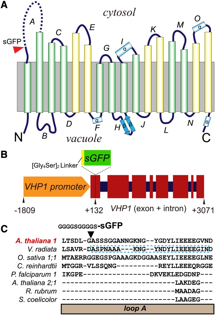
(A) Membrane topology of Vigna radiata H+-PPase (VHP1) on the basis of 3D structure (Lin et al., 2012). The insertion site of sGFP is marked by a red arrowhead. The undetermined region in loop A is shown by a broken line. The inner six transmembrane domains (TMs) and outer 10 TMs in the 3D structure of VHP1 are shown as green and yellow cylinders, respectively. Four α-helices and two β-strands of the TMs are marked as α and β, respectively.
(B) Schematic model of a VHP1-GFP construct. Exons and introns are respectively shown as wide red squares and narrow dark-blue squares. sGFP with a [Gly4Ser]2, a flexible linker, was inserted at position +132 of VHP1. Nucleotides −1809 to +3071 of the VHP1 gene were subcloned from genomic DNA, and sGFP was inserted into the +132 bp position.
(C) Sequence alignment of loop A. The region of V. radiata VHP1 not determined by crystallography is marked by a blue-dotted-line box. The alignment was generated with the Mafft program (Katoh et al., 2002).

