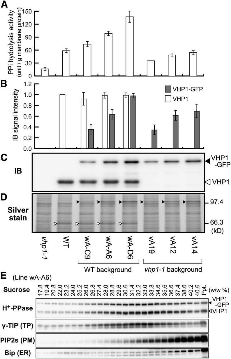
Figure 2.
Biochemical Properties of VHP1-GFP-Expressing Lines.
Crude membrane fractions (100,000g precipitate) prepared from 9-d-old seedlings of eight lines, vhp1-1, wild type (WT), three VHP1-GFP lines inserted into the wild type, and three VHP1-GFP lines inserted into vhp1-1 were assayed.
(A) PPi hydrolysis activity.
(B) Relative protein content of VHP1 and VHP1-GFP determined by immunoblotting. The amount of VHP1 in the wild type was regarded as 100%. Bars indicate sd from three independent experiments ([A] and [B]).
(C) An immunoblot with anti-H+-PPase antibody. Open and closed triangles indicate the position of VHP1 and VHP1-GFP, respectively.
(D) Silver-stained image of the crude membrane fractions (1.0 μg) after SDS-PAGE. VHP1 and VHP1-GFP are marked by open and closed triangles, respectively.
(E) Distribution of VHP1-GFP in sucrose density gradient centrifugation. Crude membrane fractions prepared from intact 3-week-old plants of wA-A6 (VHP1-GFP/wild type) were applied to a sucrose gradient (20 to 40%, w/w). After centrifugation for 18 h, the gradient was fractionated and the concentration of sucrose in each fraction determined. Distribution of H+-PPase, γ-TIP (tonoplast aquaporin), PIP2s (PM aquaporins), and Bip (ER luminal protein) was determined by immunoblotting with specific antibodies. VHP1-GFP was cofractionated with endogenous H+-PPase and a tonoplast marker, γ-TIP.

