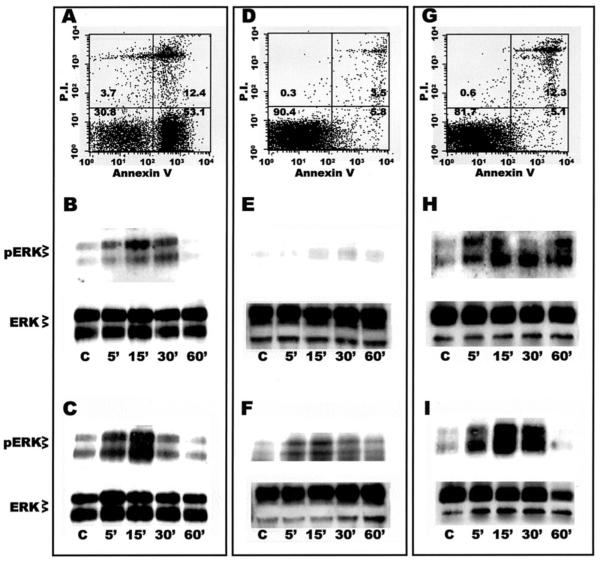
Figure 5.
Mø exposure to apoptotic or necrotic, but not viable, thymocytes rapidly activates ERK 1/2. A, D, G. Representative flow cytometric analysis of thymocyte viability. Thymocyte preparations were stained with annexin V-FITC and PI; numbers indicate the percentage of cells in each quadrant. A, thymocytes made apoptotic by 6 hour treatment with dexamethasone; D, freshly-isolated thymocytes (note virtual absence of cells showing annexin V-FITC or PI staining); G, mixture of 12% necrotic thymocytes (30 minute incubation at 56°C) and freshly-isolated thymocytes (note double staining with annexin V-FITC and PI, and virtual absence of cells staining for PI alone). B, C, E, F, H, I. Western analysis of Mø phospho-ERK (pERK) and total ERK (ERK) expression during exposure to various types of thymocytes. Resident AMø (B, E, H) and PMø (C, F, I) from normal C57BL/6 mice (4 × 10⁵ cells per well in 24 well plates) were incubated for the indicated times with 4 × 10⁶ thymocytes which were either apoptotic (B, C), freshly-isolated (E, F), or a mixture of necrotic and freshly isolated (H, I). Cytoplasmic lysates, corresponding to 400,000 Mø/lane, were electrophoresed using 12.5% SDS-PAGE run under reducing conditions, transferred by electrophoration to PVDF membranes, and immunoblotted using phospho-specific anti-ERK 1/2 Ab (top row in each panel), and then stripped and reprobed with an anti-pan ERK 1/2 Ab as a loading control (bottom row in each panel). These data are representative of two separate experiments with similar results.

