. 2014 Oct 1;3:e02935. doi: 10.7554/eLife.02935

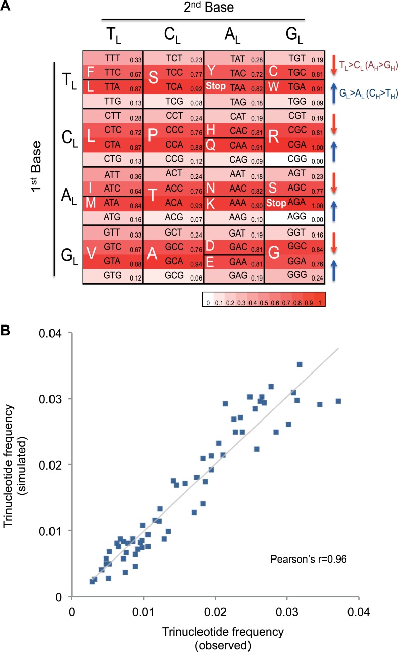
Figure 4. Mutational signature similar to processes shaping human mtDNA sequence over evolutionary time.

(A) Triplet codon depletion in human mtDNA by equivalent (CH > TH and TL > CL) mutational pressure. Relative frequency of each triplet codon within synonymous pairs (NNT–NNC or NNA–NNG) is shown by color. The arrows beside the box highlight the T > C (red) and G > A (blue) substitutional pressures on the L strand in germline mtDNA. (B) Correlation of triplet codon frequencies between from observed and from simulated evolutions of a random sequence mtDNA by the mtDNA somatic mutational signature with constraining mitochondrial protein sequences.

DOI: http://dx.doi.org/10.7554/eLife.02935.014

Figure 4.

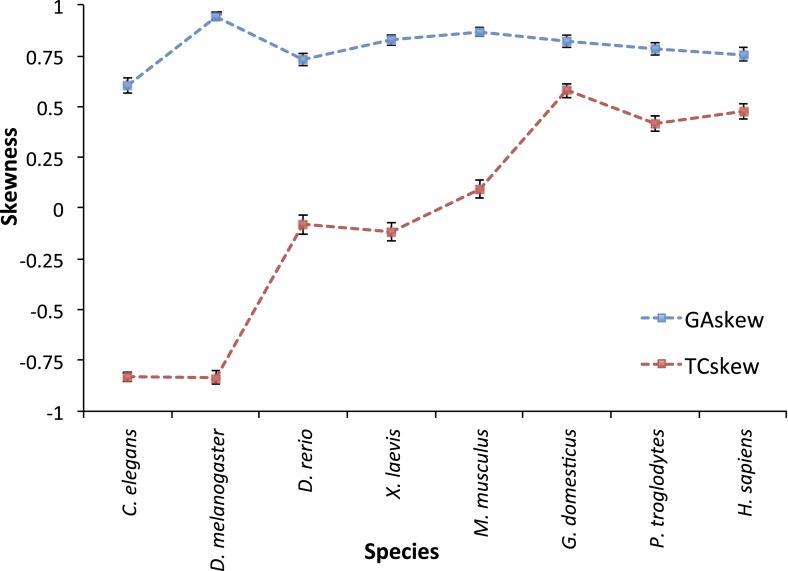
Figure 4—figure supplement 1. TC and GA skew for L strand mtDNA genes across 8 animal species.

Figure 4—figure supplement 1.

C. elegans (a nematode) and D. melanogaster (fruit fly) mtDNA appears to have GL << AL (due to CH > TH mutational pressure) and CL >> TL (due to CL > TL mutational pressure) in the third base of triplet codon in L strand genes. Therefore they seem to have predominant C > T mutational pressure without strand bias. D. rerio (zebrafish), X. laevis (frog), and M. musculus (mouse) present GL << AL (due to CH > TH mutational pressure), but similar number of CL and TL. Therefore, mtDNA of these sequences is thought to have CH > TH, with strand bias. The existence of TL > CL is not clear. Finally, mtDNA of H. sapiens, P. troglodytes (Chimpanzee), and G. domesticus (Chicken) shows clear CH > TH and TL > CL as mentioned in the main manuscript. Interestingly, TL > CL seems to be slightly stronger in the mitochondria of chicken than that of human (or chimp). We suggest there would be some differences in the mechanism of mtDNA replication across the evolution tree.
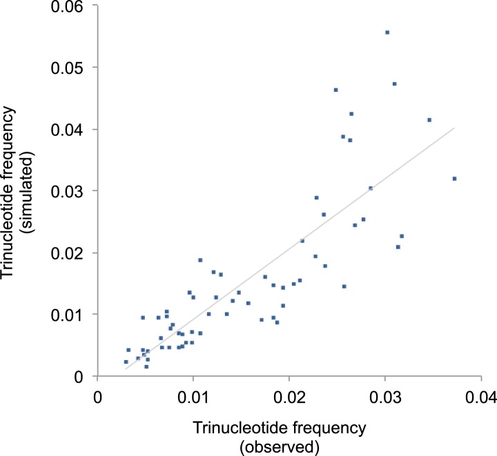
Figure 4—figure supplement 2. Correlation of triplet codon frequencies between from observed and from simulated evolutions under the mtDNA somatic mutational signature.

Figure 4—figure supplement 2.