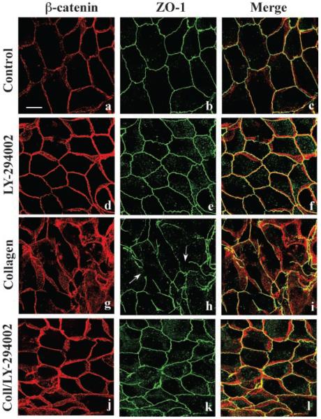
Fig. 1.
PI3-kinase regulation of adherens and tight junction structure during epithelial remodeling. MDCK cells were incubated for 6 h with DMEM (a–c; Control), DMEM containing 20 μM LY-294002 (d–f), collagen gel overlays (g–i), or collagen gel overlays containing LY-294002 (j–l). Methanolfixed cells were double-labeled for β-catenin (red staining) and ZO-1 (green staining) to visualize adherens and tight junctions, respectively, and examined by confocal microscopy. Merged 3-D projection images through the cell monolayer demonstrate that adherens and tight junctions reside adjacent to each other at the interface of the apical and basolateral membrane (Merge). Incubation with collagen gel overlays induced cell rearrangement and tight junction fragmentation (g–i; arrows) disrupting the normal staining pattern. Inclusion of LY-294002 in the collagen gels inhibited cell rearrangements and control (a–c) cell junction staining patterns were observed (j–l). Scale bar, 10 μm.

