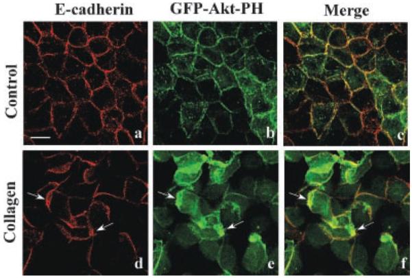
Fig. 3.

Spatial regulation of PI3-kinase. MDCK cells stably expressing GFP-Akt-PH were incubated for 6 h with DMEM (Control) or collagen gel overlays. Confocal microscopy demonstrated that GFP-Akt-PH (green staining) and E-cadherin (red staining) are localized to the basolateral membrane (a–c). After incubation with collagen gel overlays, epithelial remodeling had occurred as demonstrated by the formation of numerous lamellipodia (d–f). Under these conditions GFP-Akt-PH and E-cadherin are localized to the lamellipodial leading edge (d–f, arrows). Scale bar, 10 μm.
