Fig. 9.

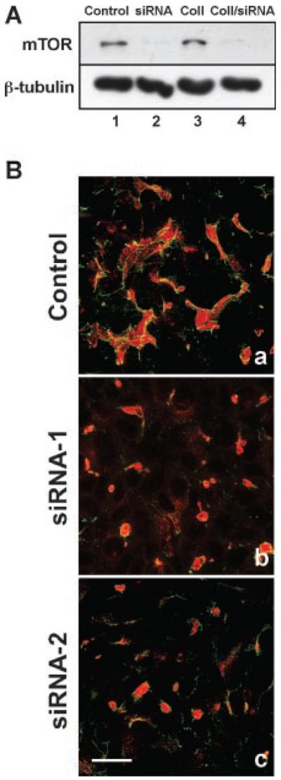
RNAi down-regulation of mTOR inhibits epithelial tubule formation. A: Western blot analysis of mTOR down-regulation. MDCK cells were incubated for 48 h with mTOR siRNA followed by 24 h incubation with, or without, collagen gel overlays. Control (lane 1) and collagen gel (Coll) treated cells (lane 3) cells were incubated with medium containing mTOR siRNA with a scrambled sequence. For mTOR down-regulation, cells were incubated in either medium (lane 2) or with collagen gel containing mTOR siRNA (lane 4). Our data demonstrate that mTOR-siRNA induced a significant down-regulation of mTOR in the presence, or absence, of collagen gel overlays (lanes 2, 4). β-tubulin was used as a protein loading control. B: Morphological analysis of epithelial tubule formation. Control cells incubated with scrambled siRNA formed elongated, multicellular tubules in collagen gel (a) identical to those lacking the scrambled RNA. However, inclusion of mTOR siRNA-1 (b) or mTOR siRNA-2 (c) in the collagen gel overlays inhibited elongated tubule formation, only allowing the biogenesis of small lumens that stained positive for gp135 (b, c; red staining) with associated ring-like tight junctions containing ZO-1 (green staining). Scale Bar, 8 μm.
