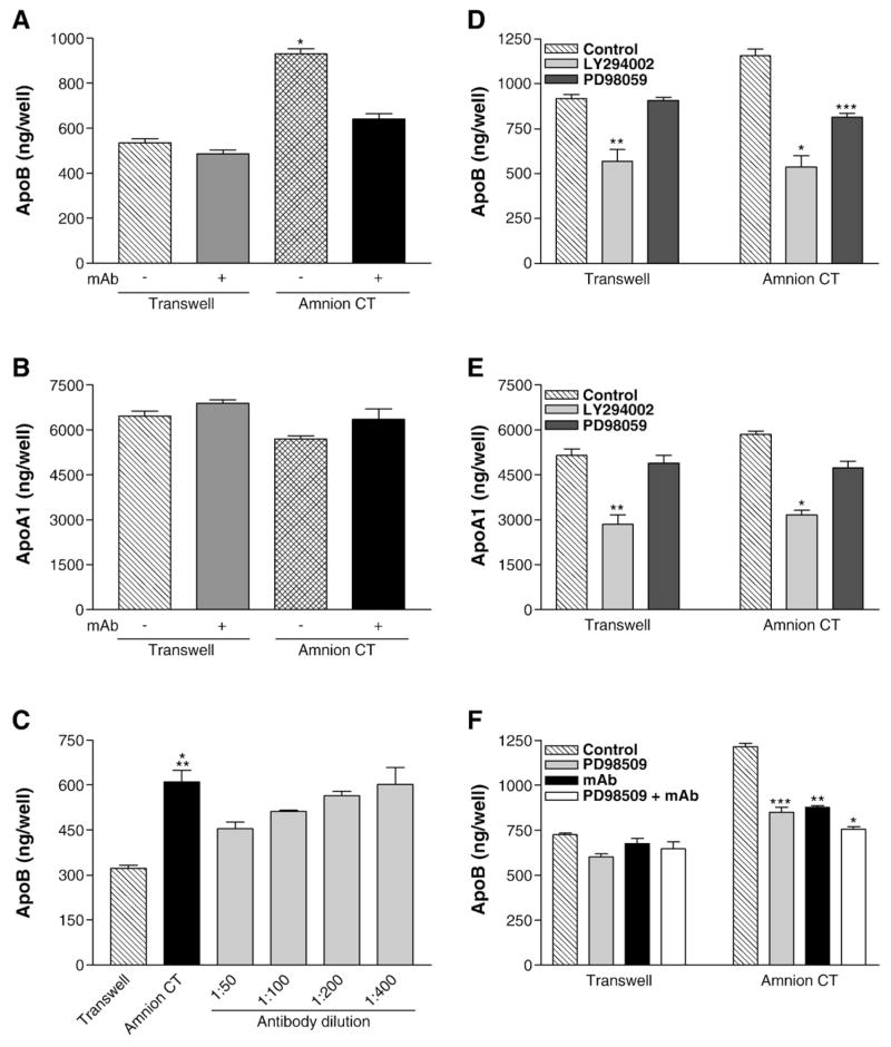
Fig. 4.
Effect of anti-integrin β1 monoclonal antibody and/or protein kinase inhibitors on the secretion of apoB and apoA1 by Caco-2 cells. Caco-2 cells were cultured for 14 days on uncoated Transwell® filters or filters coated with on HACT (Amnion CT). The apical and basal media were supplemented with fresh DMEM with or without mAb AIIB2, an anti-functional integrin β1 antibody. After 18 h, basal medium was collected and immediately assayed by ELISA for apoB (A, *p<0.0001 Amnion CT vs Transwell with or without mAb AIIB2 or Amnion CT+mAb AIIB2) or apoA1 (B). Average±s.d. values are depicted. The data are from one experiment performed in quadruplicate and are representative of four independent experiments. Panel C shows a separate experiment in which cells were incubated with different indicated dilutions of the monoclonal antibody. Average±s.d. values are plotted (*p<0.0004 Amnion CT vs Transwell, **p<0.03 Amnion CT vs Amnion CT+1:50 mAb, n=4). Panels D–F: The apical and basal media were exchanged with fresh DMEM with or without protein kinase inhibitors LY294002 (25 μM) or PD98059 (50 μM) and/or mAb AIIB2. Basal medium was collected after 18 h and immediately used to quantify apoB (D, *p<0.006, **p<0.009 Control vs LY294002, ***p<0.005 Control vs PD98059, F, *p<0.0003, **p<0.004, ***p<0.008) and apoA1 (E, *p<0.001, **p<0.008 Control vs LY294002). Average±s.d. values are depicted (n=3).

