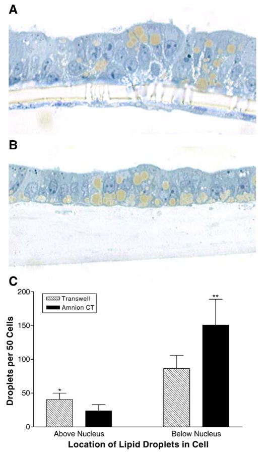
Fig. 5.
Effect of HACT on cell morphology and intracellular lipid droplets. Caco-2 cells were cultured for 14 days on uncoated Transwell® filters, or on HACT (Amnion CT). The cultures were treated for 18 h with oleic acid:taurocholate (1.6:0.5 mM), and then fixed in 2.5% glutaraldehyde and embedded for sectioning. Cross-sections show typical growth on uncoated Transwell® filters (A), or on HACT (B). Lipid droplets were counted for 50 consecutive cells on each substrate according to their location within each cell (above or below the nucleus, C). Average±s.d. are depicted (*p<0.05, **p<0.03, n=4).

