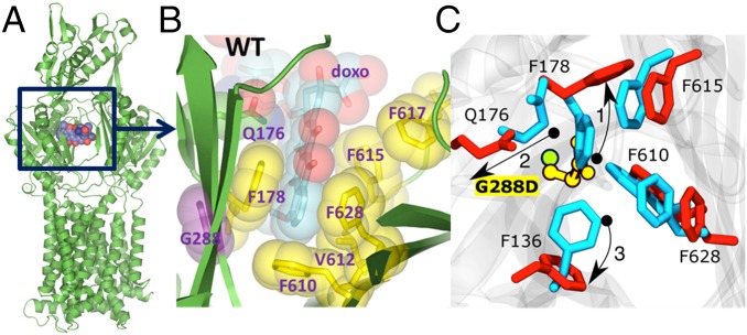
Fig. 1.
Binding of doxorubicin to the AcrB monomer. (A) Overall view of the AcrB monomer bound to doxorubicin (shown in space fill) as per PDB ID code 4DX7 (22). (B) A close-up view of the doxorubicin binding pocket in the PDB ID code 4DX7, highlighting the principal residues involved in the drug binding. (C) Close view of the binding site where important residues are shown (blue, wild type; red, mutant). Owing to the presence of the side chain of the aspartate residue in the G288D mutant, the side chain of F178 tilts away in the G288D mutant compared with its orientation in the wild-type protein (denoted by the arrow and labeled as 1). In turn, the side chain orientation of the residue Q176 is also altered in the mutated protein (denoted by 2) with respect to the wild-type form. Finally, the side chain of F136 also changes orientation (denoted by 3).

