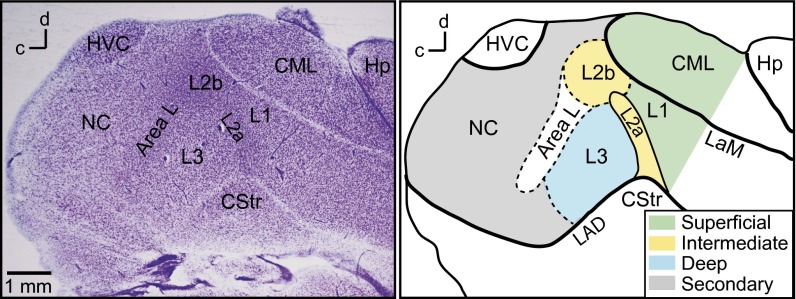
Fig. 1.
Regions of the songbird auditory cortex. (Left) Nissl-stained parasagittal section showing cell bodies and laminae. (Right) Drawing of the same section with regions colored and labeled. Dashed lines indicate boundaries that are defined by transitions in cytoarchitecture. Field L regions L1, L2, L3, and lateral caudal mesopallium (CM) form the primary auditory cortex in birds (13, 14). Field L2a and L2b are the intermediate (thalamorecipient) regions. Field L1 and CML are the superficial regions, and field L3 is the deep region. Caudal nidopallium (NC) is a secondary auditory area. Area L (13) has cytoarchitecture similar to L2b but is not known to receive thalamic input. CStr, caudal striatum; Hp, hippocampus; HVC, (proper name) a song system premotor nucleus; LAD, lamina arcopallialis dorsalis; LaM, lamina mesopallialis.

