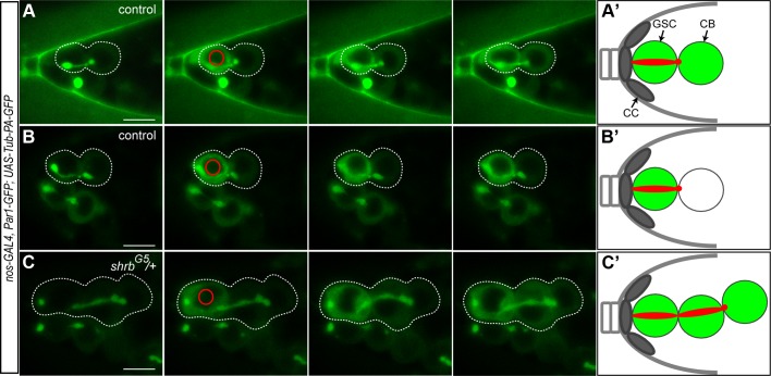
Figure 5. The cells of the stem-cyst share the same cytoplasm.
Selected time points of live imaging experiments performed on germaria expressing UAS-Par1-GFP and UAS-Tubulin-PA-GFP under the nos-GAL4 promoter. Tub-PA-GFP is photoactivated in the region defined by the red circle in the GSC, and the fluorescence diffusion to the neighboring cells is observed. (A and B) WT female GSCs in mid-G2 phase. (A) Abscission between the GSC and the CB did not yet occurred as the GFP diffuses to the CB. (B) GFP does not diffuse to the CB. Abscission has occurred and the cells no longer share their cytoplasm. (C) Stem-cyst of shrbG5/+ female. After photoactivation in the GSC, the GFP diffuses through the 2 neighboring cells. This shows that the cells of the stem-cyst do not complete abscission and stay connected, sharing their cytoplasm. Dotted lines surround: GSC/CB pair (A and B), stem-cysts of 3 cells (C). (A’, B’ and C’) Schematic representation of the Tubulin-PA-GFP diffusion from the GSC to the CB (A’ and B’) or within a stem-cyst (C’). Scale bar: 10 μm.

