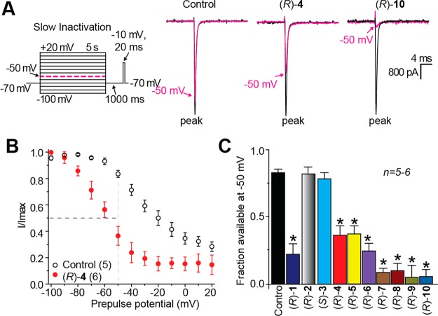
Figure 6.
Effects of the chimeric compounds ((R)-1, (R)-4–(R)-10) and the parent compounds ((R)-2, (S)-3) on inactivation state of Na+ currents in rat embryonic cortical neurons. (A) Inactivation voltage protocol. Currents were elicited by 5 s prepulses between −100 and +20 mV (in 10 mV increments), and then fast-inactivated channels were allowed to recover for 1000 ms at a hyperpolarized pulse to −70 mV before testing for the fraction of available channels for 20 ms at −10 mV. Finally, the fraction of channels available at −10 mV was analyzed. Representative current traces from cortical neurons in the absence (control, 0.1% DMSO) or presence of 10 μM (R)-4 or (R)-10 are illustrated. The black and pink traces represent the peak current evoked (between −100 to −80 mV and −50 mV, respectively (also highlighted in the voltage protocol as a dashed pink line). (B) Summary of steady-state activation curves for neurons treated with 0.1% DMSO (control) or 10 μM (R)-4. For compounds that mediate inactivation, (R)-4 shown, significant enhancement of inactivation is evident by separation of the curves starting at −80 mV. (C) Summary of the fraction of current available at −50 mV for neurons treated with 0.1% DMSO (control) or 10 μM of the indicated compounds. Asterisks (*) indicate statistically significant differences in fraction of current available between control (0.1% DMSO) and the indicated compounds (p < 0. 05, Student’s t test; n = 5–6 cells per condition).

