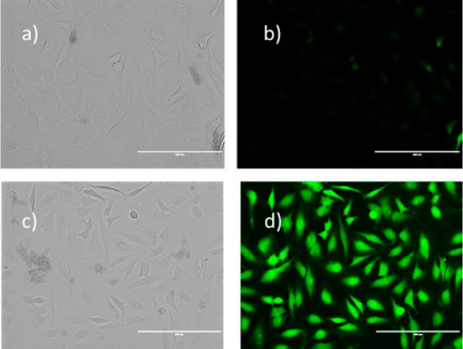
Figure 5.

Fluorescence images of H2S in HeLa cells using SeP2. Cells were incubated with the probe (50 μM) for 30 min, then washed, and subjected to different treatments. (a and b) Control (no Na2S was added); (c and d) treated with 100 μM Na2S (scale bar: 100 nm).
