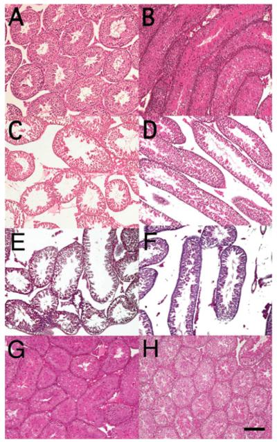
Figure 4. Histological abnormalities of Pla2g4a−/− testes.
Histological analysis of testes from juvenile 44-day-old Pla2g4a+/+ (A and B) and Pla2g4a−/− (C–F) mice, and fully mature 119-day-old Pla2g4a+/+ (G) and 122-day-old Pla2g4a−/− (H) mice. Representative cross-sections (A, C, E, G and H) and parallel sections (B, D and F) of haematoxylin and eosin-stained seminiferous tubules are shown. Juvenile wild-type testes show a highly integrated structure of seminiferous tubules with normal spermatogenesis within them. In the testes of juvenile Pla2g4a−/− mice, elongated spermatids were also seen. However, the structure of the seminiferous epithelium tends to be disorganized and loose and disrupted junctions are evident among tubules. Pla2g4a−/− testes (H) as well as Pla2g4a+/+ testes (G) of approximately 120-day-old mice appear histologically normal. Scale bar is 100 μm and is applicable for all panels.

