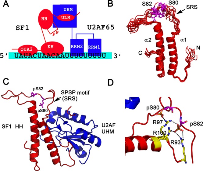
Figure 2.

X-ray crystallographic and NMR structures of phosphorylated SF1. (A) Schematic showing the domain organization and interactions between SF1, U2AF, and the RNA. (B) Solution NMR structural ensemble (PDB code 2M09) of the free helix hairpin (HH) motif consisting of two α-helices in an antiparallel arrangement connected by a flexible linker is shown. The serines that are phosphorylated are in magenta in a dynamic SPSP loop. (C) The 2.29 Å crystal structure of phosphorylated SF1 HH bound to the U2AF UHM (PDB code 4FXW) is shown. SF1 is in red, and U2AF in blue. The phosphorylated serines are depicted in magenta. (D) Interactions of the phosphates with neighboring arginines to form an “arginine claw” are shown.
