
Figure 5.
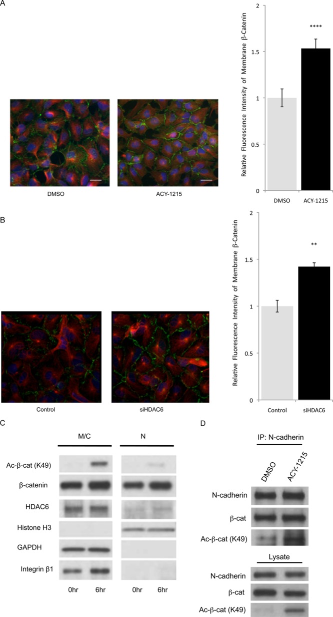
Effect of HDAC6 inhibition on membrane localization of β-catenin. (A, B) Immunofluorescence staining for β-catenin treated with HDAC6 inhibitor (A) and HDAC6 siRNA (B) in human NPCs. Cells were treated with 5 μM ACY-1215 or DMSO for 18 h (A) or with HDAC6 siRNA and control siRNA for 72 h (B) and imaged with anti-β-catenin antibody (green), anti-β-tubulin antibody (red), and Hoechst 33342 (blue). Scale bar, 20 μm. Quantification of β-catenin levels at the membrane represent two independent experiments with three fields of view each at 20× magnification. Error bars indicate standard deviation. ** and **** denote significance at p < 0.01 and p < 0.0001, respectively (unpaired t test). (C) Immunoblot analysis for nuclear (N) and membrane/cytoplasmic (M/C) fractions for NPCs treated with HDAC6 inhibitor ACY-1215 (5 μM) or DMSO for 6 h. Histone H3 is shown as a nuclear marker, GAPDH as a cytoplasmic marker and integrin β1 as a membrane marker. (D) Immunoblot analysis for Ac-Lys49-β-catenin bound to N-cadherin. Human NPCs were treated with ACY-1215 (5 μM) or DMSO for 24 h, lysates immunoprecipitated with anti-N-cadherin antibody and immunoblotted with antibodies against β-catenin and Ac-Lys49-β-catenin.