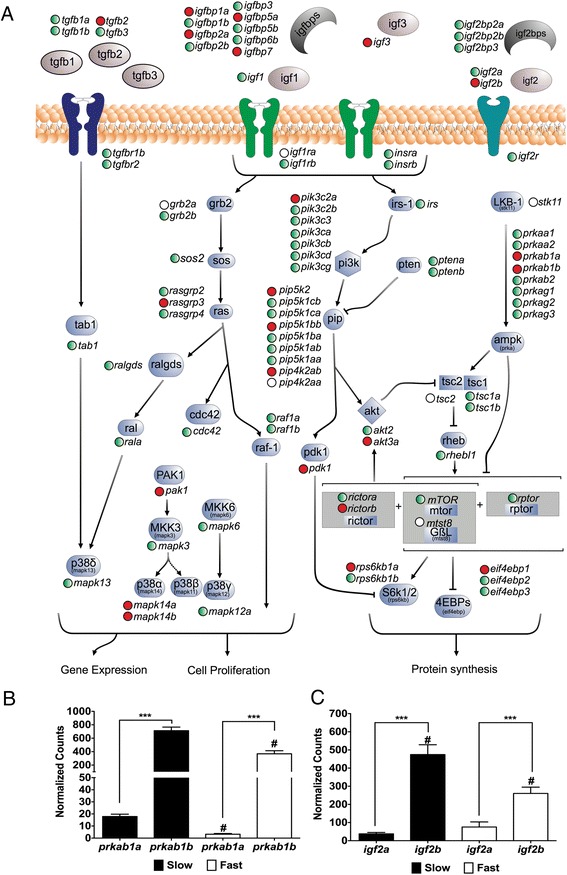
Figure 2.

Digital gene expression of the Pi3k/mTor pathway components in pacu fast and slow skeletal muscle. (A) Pi3k/mTor components represented in the transcriptome mapped into a reconstruction of the same pathway. “Red circles” and “Empty circles” indicate components significantly higher in slow and fast skeletal muscle respectively muscle (FDR < 0.05). “Green circles” indicate components with no significant differences between fibre types. (B) igf2a and igf2b digital gene expression. (C) prkab1a and prkab1b digital gene expression. Digital gene expression is represented using a logarithmic scale for slow (full bars) and fast (empty bars) skeletal muscle. Significant differences for paralogues within (***; FDR < 0.001) and between (#; FDR < 0.05) fibre types are indicated. Values represent mean ± SE (n = 5). Igf2: insulin-like growth factor 2; Prkab1: 5’ AMP-activated protein kinase subunit beta 1.
