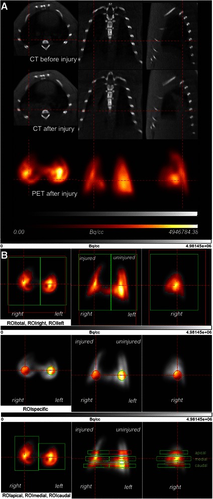
Figure 2.

Example of image processing after co-registration. (A) Co-registered PET and CT images in the transverse, coronal, and sagittal planes of one animal; the CT image before and after injury and the corresponding PET scan image are shown. Dark areas in the CT scan represent aerated zones. The areas of high density were formed in the right lung after injury (red lines). The PET scan shows the distribution of PBF in the left and right lungs. (B) PET images and definition of the ROI for one lung in the transverse, coronal, and sagittal planes. The tracer activity in the lung is indicated in grayscale, from lowest (black) to highest activity (white). ROI masks were used for the threshold analysis and to calculate the median maximum standardized uptake values (SUVmax) for each side of the lung in a 3-dimensional environment. The thresholds in the ROI masks for the entire lung (red: ROItotal), the left and right lung (green), the spherical masks (ROIspecific) (0.24 cm3) and the 6 ROIs (apical, medial, and caudal) are indicated by the highlighted regions of the color-coded areas. Color coding shows a relative scale in which the measurements are normalized to their mean values and represent regional tracer concentrations of Ga-68 microspheres from high (white) to low (black), as determined by PET in Bq/cm3. Red lines are focused on the area of injury.
