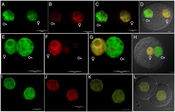
Figure 8.

Immunofluorescent labeling of H4K12ac and 5-methylcytosine (5mC) in mouse early embryos. Double stained embryos with anti-H4K12ac antibody (green) (A, E, I), anti-5mC antibody (red) (B, F, J), merged (C, J, K), merged with DIC (D, H, L). (A-D) Pronucleus at stage PN3. (E-H) Pronucleus at stage PN5. (I-L) Two-cell embryo. Maternal pronucleus (♀) and paternal pronucleus (♂). Scale bar represents 20 μm.
