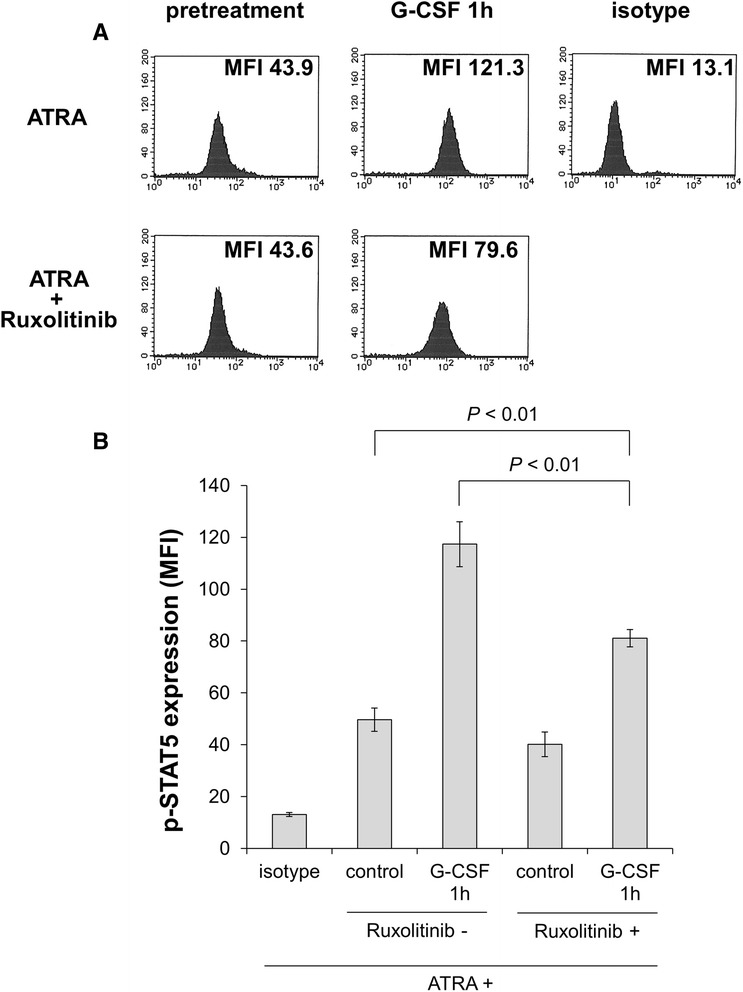
Figure 5.

After treatment with 100 nM ATRA with or without 320 nM ruxolitinib for 5 days, HT93A cells were stimulated with 50 ng/mL G-CSF for 60 min. Expression of phosphorylated STAT5 was evaluated by intracellular staining and analyzed by flow cytometry. Ruxolitinib-treated HT93A cells showed significantly lower phosphorylated STAT5 expression. Histograms (A) and bar graphs (B) represent phosphorylated STAT5 expression. Experiments were independently repeated three times and results are shown as mean ± SD.
