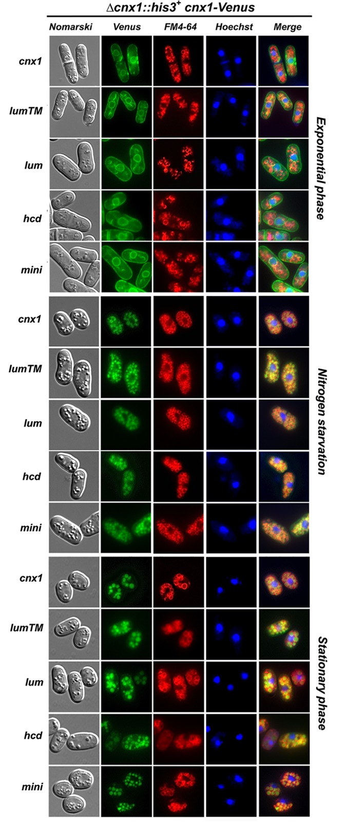
Fig 6. The processing and the ER-to-vacuole trafficking of calnexin and its mutant versions occur under nitrogen starvation and in stationary phase.

Subcellular localization of calnexin-truncated mutants fused to Venus. Strains cnx1::his + + pREP41lumenalTM_cnx1-Venus (SP19207), pREP41lumenal_cnx1-Venus (SP19174), pREP41Δhcd_cnx1-Venus (SP19211), pREP41mini_cnx1-Venus (SP19212) and pREP41cnx1p-Venus (control, SP19201) were grown in EMM to mid-logarithmic phase. The culture was split into two, half was maintained until stationary phase for 3 days, and the other half was shifted to EMM-N medium to induce nitrogen starvation (24h). The cells were analyzed by fluorescence microscopy for Venus for the localization of Cnx1-Venus), FM4–64 was used as a marker for the vacuole, and Hoechst 33342 was used as a marker for the nucleus. Nomarski bright-field microscopy was used to monitor cell morphology. Merged images were used to determine colocalization.
