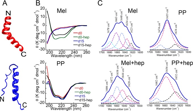
Fig 1. Structural characterization of Mel and PP.
(A) Structural model of Mel (red, PDB ID: 2MLT) and PP (blue, bovine PDB ID: 1BBA). (B) CD spectra of Mel and PP at day 0 (d0) and after 15 days (d15) in presence and absence of heparin. After the addition of heparin and subsequent incubation for two weeks, the secondary structure of PP remained mostly unchanged (helical). (C) FTIR spectra of two weeks incubated PP and Mel (in the absence and presence of heparin). Y-axis represents the absorbance (AU) and X-axis represents the wavenumber (cm-1). Wavenumbers corresponding to the maximum absorbance are represented with arrow marks. Consistent with CD data, FTIR study also showed that in the presence of heparin, unstructured Mel transformed into helical conformation, whereas PP remained mostly helical both in presence and absence of heparin after incubation.

