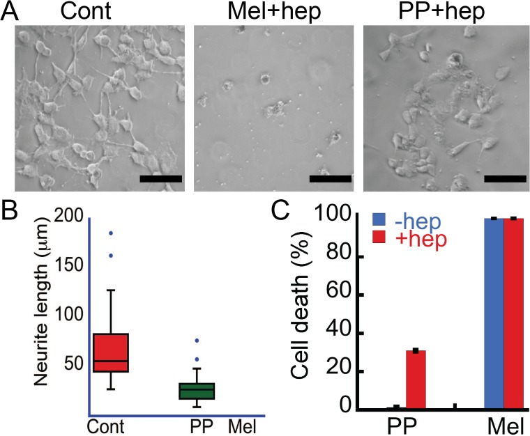
Fig 7. Cytotoxicity of Mel and PP oligomers.
(A) Phase contrast images of SH-SY5Y cells showing damaged morphology of cells by PP and Mel oligomers. Scale bars are 100 μm. (B) Neurite length of SH-SY5Y cells treated with Mel and PP oligomers that were quantified using ImageJ software (NIH). The cells treated with oligomers showing reduced neurite length when compared to control. Due to complete death of cells in Mel oligomer treated samples, the calculation was only conducted for control samples (buffer) and PP oligomers treated samples. (C) LDH assay depicting % cell death in SH-SY5Y cells treated with oligomers. Both PP and Mel oligomers (formed in the presence of heparin) showed cytotoxicity. PP incubated in the absence of heparin did not show any toxicity; however, Mel incubated alone showed cytotoxicity.

