Fig. 4.
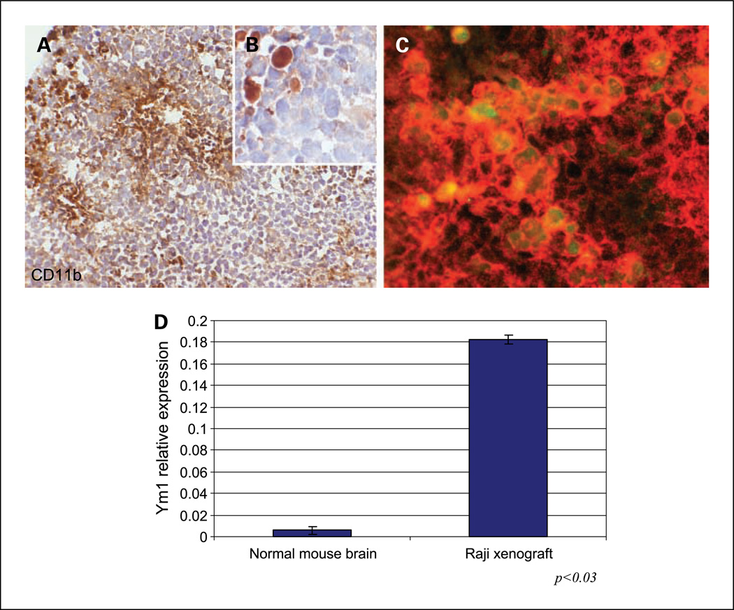
Evidence of macrophage recruitment and M2 polarization in intracerebral Raji xenograft model. A, micrograph of CD11b immunostaining shows abundant foci of macrophage infiltration within highly cellular CNS lymphoma xenograft (hematoxylin counterstain, ×200). B, higher magnification shows specificity of CD11b immunoreactivity by tumor macrophages with absent staining by lymphoma cells (×1,000). C, fluorescently labeled antibodies were used to show the presence of intratumoral macrophages using CD11b (Alexa Fluor 594). M2 macrophages were positive for CD11b and Ym1 (Alexa Fluor 488). The merged image indicates M2 macrophages exhibiting cytoplasmic expression of Ym1 by CD11b+ macrophages. D, quantitative RT-PCR was used to confirm increased RNA levels of mouse Ym1 associated with intracerebral lymphoma growth.

