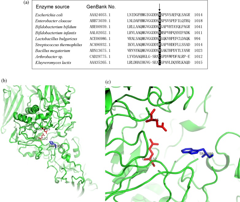
Fig 1. Multiple alignment of GH2 β-galactosidases (a) and the homology model of the whole BgaL3 (b) and its enlarged active center (c).
The amino residues in the black boxing region are parallel to the E. coli W999. The putative catalytic residues of BgaL3, Glu465 and Glu532, locate at the bottom of the active center (in red), while the crucial residue involved in substrate specificity, W980, stands at the entrance of the active center (in blue).

