
Fig 2. Example run of the compressive test procedure for one skin specimen when varying strain rates.
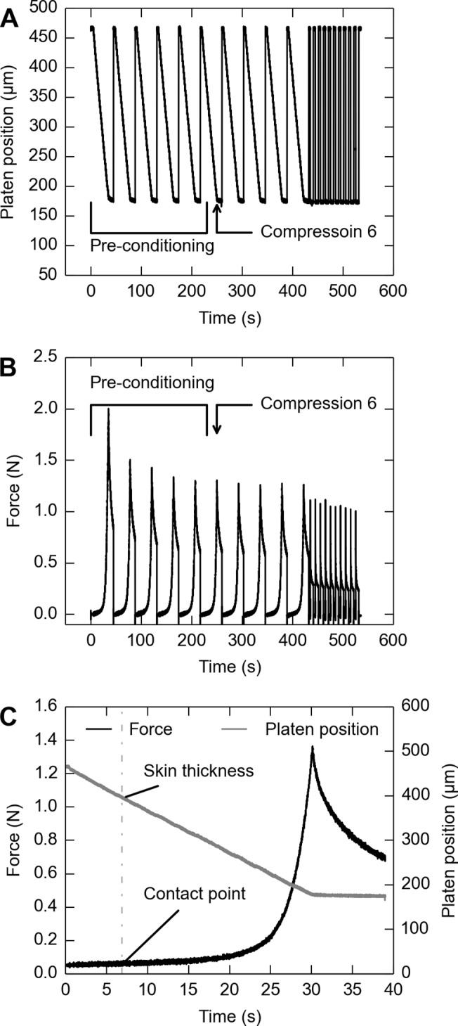
A: Position of the compression platen over time, as measured by its distance from the fixed platform. B: Reaction force at the compression platen. C: Magnified view of reaction force and platen position for Compression 6, demonstrating that “skin thickness” was defined by “contact point” as determined from the force trace. The platen was moved into the skin with an acceleration of 0.06 s−1 for each of the first 10 repetitions. Then, 10 additional compressions were performed at 22.88 s−1. The 6th compression was analyzed in each sequence of 10 compressions.