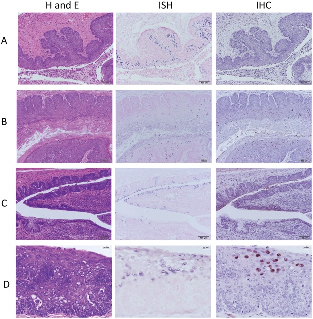
Fig 2. A-D H and E, in situ hybridization and immunohistochemistry at four time points.
A) Time point 9 weeks post infection. Vaginal lesions; ISH positive cells greatly outnumber IHC positive cells. 20X B) Time point 14 weeks post infection. Vaginal lesions exhibit greatly increased IHC signal. 20X C) Time point 25 weeks post infection. Endocervical canal and dorsal fornix. ISH positive squamous and mucous cells are present in the caudal canal. Only squamous cells are IHC positive. 20X D) Time point 30 weeks post infection. Cytologic atypia in the stratified squamous vaginal epithelium, 40X.

