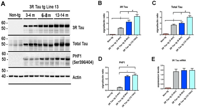
Fig 2. Effects of aging on biochemical alterations in the higher expresser mutant 3R Tau tg mice.

A Representative Western blot (SDS) and analysis of levels of B 3R Tau, C Total Tau, and D PHF-1. Across all antibodies there was a significant increase in the amount of protein at the older time points (12–14 months of age) compared to the earliest time point (3–4 months of age) and non-tg mice. E. Levels of 3R Tau mRNA levels at 3–4, 6–8, and 12–14 months of age were uniformly increased compared to non-tg mice. N = 8 for each age group * = P < 0.05 when compared to non-tg control using one way ANOVA with Dunnett’s posthoc test. # = P < 0.05 when comparing Line 2 and 13 using one way ANOVA with Fisher’s post hoc test.
