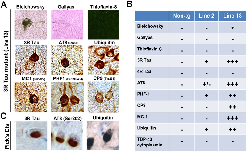
Fig 8. Histochemical and immunocytochemical characteristics of the aggregates in the higher expresser mutant 3R Tau tg mice.
A. Representative photomicrographs (frontal cortex) of brain sections from the higher expresser Line 13 mice stained with Bielchowsky and Gallyas silver impregnation technique and Thioflavin-S and immunostained with the antibodies against 3R Tau, pTau (AT8, CP9, MC-1, PHF-1) and ubiquitin. B. Summary table with semiquantitative assessment of the abundance of pathological Tau lesions in the brains of the mutant 3R Tau tg mice. C. Representative photomicrographs of frontal cortex from a Pick’s disease case immunostained for 3R Tau, AT8, and ubiquitin. Scale bar = 10 μm. Mice were 8–10 months old from each line.

