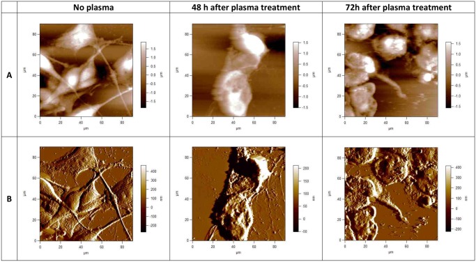
Fig 2. AFM images of fixed human brain glioblastoma (U87) cells, taken using Igor Pro 4 90 x 90 μm scans.
Upper row (A), topographies of 2-dimensional images of U87 cells before and 48h and 72h after plasma treatment; lower row (B) corresponding deflection signal images. Cells were treated with cold plasma for 30 s. All images presented here were obtained in contact mode at room temperature and scanned in PBS.

