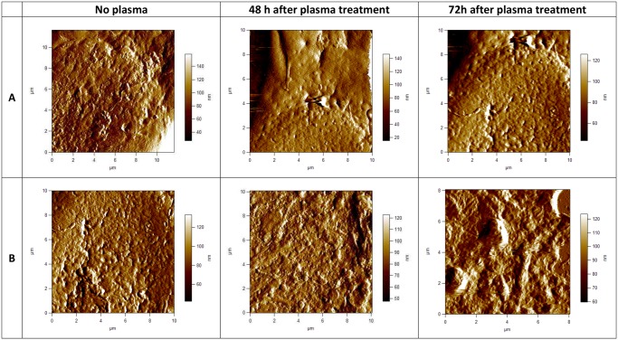
Fig 3. AFM images of cell membrane surface of fixed human astrocytes (E6/E7) and fixed human brain glioblastoma (U87) cells.
Scan size is approximately 10x10 μm. In the upper row (A), 2-dimensional deflection signal images of cell membrane surface of E6/E7 cells are shown and in the lower row (B) 2-dimensional deflection signal images of cell membrane surface of U87 cells. All images presented here were obtained in the contact mode at room temperature and scanned in PBS.

