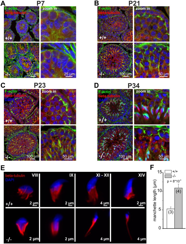
Fig 4. Cytoskeletal defects in GBA2 knockout-testis already occur during the first spermatogenic wave.
(A) Immunofluorescent labeling of the cytoskeleton in wild-type (+/+) and GBA2 knockout-testis (-/-) at P7. Microtubules have been labeled using an anti-beta tubulin III antibody (red), F-actin using Alexa Fluor Phalloidin 488 (green), and the DNA using DAPI (blue). Scale bars are indicated. (B) See (A) for P21. (C) See (A) for P23. (D) See (A) for P34. (E) Development of the manchette in spermatids. The manchette was stained with beta-tubulin (red), DNA was labeled with DAPI (blue). Different developmental stages are indicated. (F) Manchette length. The manchette length of spermatids from wild-type (+/+) and GBA2 knockout-mice (-/-) was determined using ImageJ. At least 30 cells have been analyzed per genotype. Data are shown as mean ± S.D.; n numbers and p values calculated using One-Way ANOVA are indicated.

