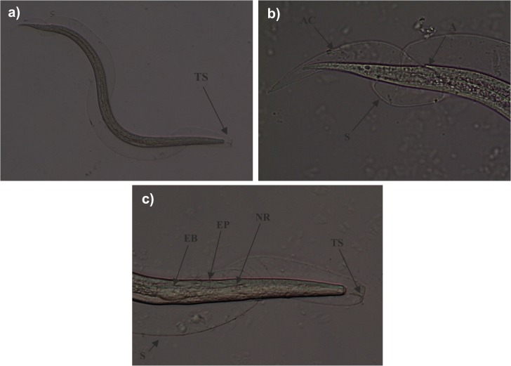
Fig 4. Light microscopy of third stage larvae (L3) morphologically compatible with Angiostrongylus cantonensis, obtained from Theba pisana.
General view of third-stage larvae with one sheath (S). A characteristic “T”-shaped structure (TS) is apparent at the anterior end of the sheath that surrounds the L3 larva (a). Posterior end of L3 larva with anus (A) at subterminal position. Anus cuticle (AC) is completely molted and can be seen on the sheath (b). Anterior end of L3 larva with esophagus bulbus (EB), excretory pore (EP) and nervous ring (NR) (c).

