Figure 1.

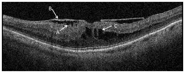
Example of a B-scan with an epiretinal membrane consisting of corrugation of the retinal surface (A) with bridging of the membrane (B) across the top of the corrugation. A cluster of cysts (C) is visible in the outer retinal layers.

Example of a B-scan with an epiretinal membrane consisting of corrugation of the retinal surface (A) with bridging of the membrane (B) across the top of the corrugation. A cluster of cysts (C) is visible in the outer retinal layers.